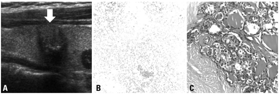
Fig. 2.
A 50-year-old female with papillary thyroid carcinoma. (A) Ultrasonography shows well-circumscribed and isoechoic nodule (arrow) with a shape that is taller than it is wide and internal microcalcification in the thyroid gland; this nodule was considered suspicious. (B) Fine-needle aspiration cytology was interpreted as adenomatous hyperplasia based on the presence of flat sheets of follicular cells in the bloody background (Giemsa stain ×200 original magnification). Because of the discordant results between the sonographic findings and the cytological results, surgical excision was performed. (C) Thyroidectomy specimen shows a typical papillary carcinoma nucleus with a few grooves, clearing and pseudoinclusions, compatible with papillary thyroid carcinoma (hematoxylin and eosin (H&E) ×400 original magnification).

