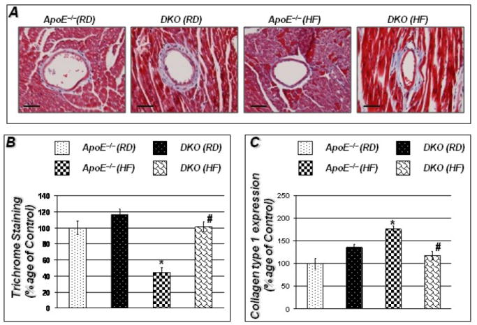
Figure 3. HF diet promotes collagen degradation in hearts of ApoE−/− mice; protection by PARP-1 gene deletion.

Figures 3A show trichrome staining for collagen around vessels in hearts of the experimental groups. Figure 3B represents quantitation of Trichrome staining conducted using Image-Pro Plus software and expressed percentage of control. Figure 3C shows that decreased collagen content in ApoE−/− mice was accompanied by increased collagen synthesis as determined by real time PCR for collagen type 1. cDNA was subjected to real-time PCR with primers specific to murine collagen type-1; β-actin was used as an internal control for normalization of expression values. Scale bar: 50μm P value (<0.05) *compared to ApoE−/− RD, #compared to ApoE−/− HF (16 weeks).
