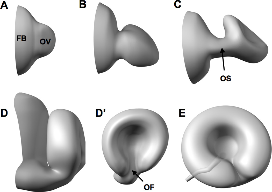
Figure 2. Development and morphogenesis of the zebrafish eye.

Eye development commences around 12 hpf as the optic vesicle (OV) evaginates from the forebrain (FB) (A). The optic vesicle then elongates into a flattened wing-like structure at around 16 hpf (B) that is attached to the forebrain through a transient structure called the optic stalk (OS in C). The eye subsequently rotates and invaginates (C) to form the ‘optic cup’ at around 24 hpf as depicted in D (anterior view) and D’ (lateral view). Morphogenesis of the embryonic eye is mostly complete by 48 hpf as the optic fissure (OF in D’) is closed and neurogenesis of the retina is underway [54, 68, 286].
