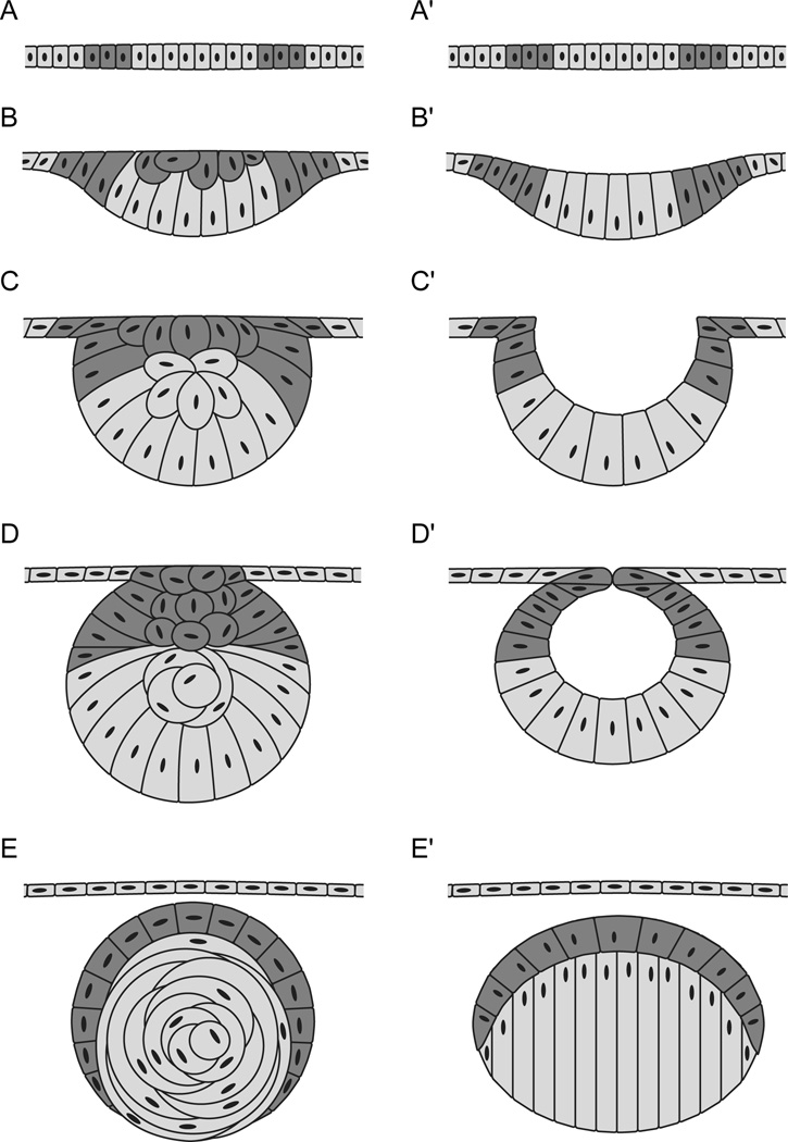
Figure 3. Early lens development in zebrafish and mouse.
After [58]. In zebrafish (A–E) and mouse (A’–E’), the surface ectoderm overlying the optic cup (A, A’) thickens to form the lens placode (B, B’). In zebrafish, the lens mass delaminates from the surface ectoderm (C, D), while in mouse the lens placode evaginates to form the lens vesicle (C’, D’). Primary lens fibers in the zebrafish elongate within the lens mass in a circular fashion (E), whereas the primary lens fibers in the mouse elongate to the anterior to fill the lens vesicle space (E’). The remaining surface ectoderm becomes the corneal epithelium in both zebrafish and mouse. [57, 59, 67, 191]. Anterior is to the left.

