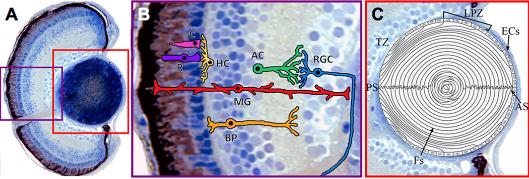
Figure 4. Structure of the zebrafish eye.
(A) Transverse histological section of a wild-type zebrafish eye at 5 dpf. (B) Illustration of the major neuronal and glial cell types in the zebrafish retina. Rods and cones relay sensory input to retinal interneurons (Horizontal and Bipolar Cells). Following synaptic interactions with Amacrine Cells, the information is passed to the output neurons, the Ganglion Cells. Müller glia perform multiple functions in the retina, including maintaining retinal health and structure (see ‘Müller Glia’ section in text). C, Cones. R, Rods. HC, Horizontal Cells. BP, Bipolar. AC, Amacrine Cells. RGC, Ganglion Cells. MG, Müller Glia. (C) Diagram of the zebrafish lens at 5 dpf. Lens epithelial cells surround the anterior periphery; proliferation of these cells is mainly restricted to the lateral proliferative zone. Epithelial cells at the transition zone exit the cell cycle, migrate, elongate, and degrade their light-scattering organelles to become new secondary fibers. Tightly-packed lens fibers which extend from the posterior suture to the anterior suture make up the bulk of the lens. [57–60, 193–195]. ECs, lens epithelial cells. LPZ, lateral proliferative zone. TZ, transition zone. Fs, lens fibers. AS, anterior suture. PS, posterior suture.

