Figure 7.
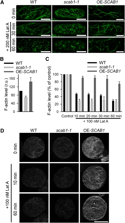
SCAB1 Stabilizes Actin Filaments in Vivo.
(A) Actin filament organization in hypocotyl epidermal cells from wild-type (WT; left column), scab1-1 mutant (middle column), and OE-SCAB1 (right column) plants expressing 35S:fABD2-GFP before and after 30 or 60 min of Lat A treatment.
(B) The relative level of F-actin in suspension cells of the wild type, scab1-1, and OE-SCAB1 was quantified before Lat A treatment. The F-actin level in the wild type was arbitrarily set to 100%. The data given are the means ± sd (n = 3). r.u., relative unit.
(C) Filamentous actin in suspension cells of the wild type, scab1-1, and OE-SCAB1 was quantified at different time points after the addition of Lat A. Each bar represents the mean value (± sd) of three independent experiments. The level of F-actin without Lat A treatment was set at 100% for each cell type.
(D) Alexa-488-phalloidin-stained MFs in suspension cells of the wild type (left column), scab1-1 (middle column), and OE-SCAB1 (right column) before and after 10 or 60 min of Lat A treatment. Scale bars, 20 μm.
[See online article for color version of this figure.]

