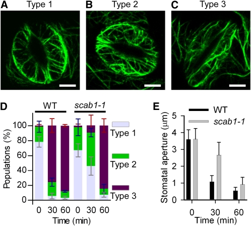
Figure 8.
The scab1 Mutation Delays Actin Reorganization during Stomatal Closure.
(A) to (C) Confocal images of guard cells in rosette leaves from Pro35S:GFP-fABD2-GFP transgenic plants in a wild-type background. Actin organization was classified into three groups. Representative images of type 1, radial array (A); type 2, random meshwork (B); and type 3, longitudinal array (C) are shown. Scale bars, 5 μm.
(D) Histograms showing the actin organization in guard cells from wild-type (WT) and scab1-1 plants at the indicated times after abscisic acid treatment. The guard cells were classified into three groups: type 1, type 2, and type 3. The data represent the mean ± sd of six independent experiments; 100 guard cells per line were measured at the indicated times.
(E) Stomatal aperture in wild-type and scab1-1 mutant leaves at the indicated times after abscisic acid treatment. The data represent the mean ± sd of three independent experiments. At least 50 stomata were analyzed per line.

