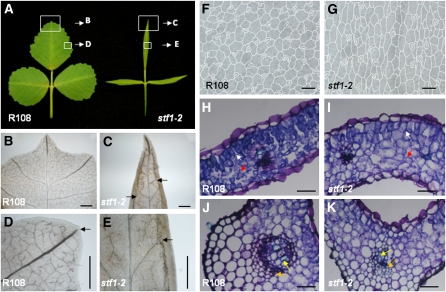
Figure 2.
The stf Mutant Is Severely Defective in Leaf Vascular Patterning.
(A) Wild-type, R108, and stf1-2 mature leaves showing the regions for close-up described in (B) to (E).
(B) to (E) Leaf material observed through a light microscope after clearing with lactic acid.
(B) Major and minor veins of R108 leaf.
(C) Disorganized and poorly developed major veins in stf. Major veins are forming near the margins (one on either side of the midvein) along the proximodistal axis (arrows).
(D) R108 major vein extends close to the margin with its tip aligned to the serration and is open ended (arrow).
(E) stf major vein poorly developed and connected to marginal vein (arrow).
(F) R108 leaf epidermal cells viewed through a light microscope.
(G) Epidermal cells of stf leaf showing narrower width.
(H) Transverse section through R108 leaf blade showing palisade mesophyll (white arrow) and spongy mesophyll (red arrow) cells. Sections were stained with Toluidine Blue.
(I) Transverse section through stf leaf blade showing the poor distinction between palisade mesophyll (white arrow) and spongy mesophyll (red arrow) cells.
(J) Transverse section through the midrib of R108 leaf showing xylem (yellow arrow) and phloem (orange arrow) vessels.
(K) Transverse section through stf midrib showing poorly differentiated xylem and phloem vessels (yellow and orange arrows) and cortical tissue. Scale bars in (B) to (E) = 500 μM, in (F) and (G) = 50 μM, and in (H) to (K) = 100 μM.

