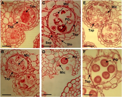
Figure 10.
The undead Mutant Exhibits Premature Tapetal Degeneration.
Sections (3 μm) of undead and wild-type anthers stained with safranin. MC, meiotic cell; Tap, tapetum; Tds, tetrads; Mic, microspores; PG, pollen grain; Sep, septum; Sto, stomium. Bars = 20 μm.
(A) Stage 7, tetrads are formed. The tapetum exhibits increased vacuolation similar to the myb80 T-DNA mutant (Li et al., 2007).
(B) Stage 8, the tapetum appears sparse, exhibiting advanced degeneration commonly seen in wild-type stage 10 anthers.
(C) Stage 10, most of the microspores are aborted. A thin tapetal layer still remains.
(D) Stage 13, anther dehiscence and the breakage of both septum and stomium layers occur as normal. The collapsed pollen grains clump together and are not able to be released.
(E) Wild-type stage 8.
(F) Wild-type stage 11.

