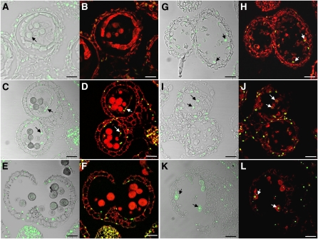
Figure 11.
Tapetal PCD Occurs Prematurely in the myb80 Mutant.
Confocal microscopy of DNA fragmentation detected using the TUNEL assay in 6-μm sections of wild-type ([A] to [F]) and myb80 ([G] to [L]) anthers. TUNEL-positive signal is indicated by the green fluorescence of fluorescein, and nuclei fluoresce deep red due to the counterstain propidium iodide. Bars = 25 μm.
(A) and (B) Wild-type anther stage 9 with the first appearance of the pollen coat (arrow) was TUNEL-negative.
(C) and (D) TUNEL signal is first observed in the tapetum of wild-type anthers at stage 10 (arrows), which correlates with the onset of tapetal cell breakdown.
(E) and (F) At stage 13, the tapetum has degenerated completely, and TUNEL signal is visible only in the anther epidermis and endothecium.
(G) and (H) myb80 anthers corresponding to stage 9 exhibit TUNEL signal in the tapetal layer but not in microspores (arrows).
(I) and (J) At stage 10 in myb80, TUNEL-positive signals appeared in the collapsing microspores (arrows).
(K) and (L) At stage 13 in myb80, TUNEL-positive nuclei are observed in the collapsed pollen (arrows).

