Figure 3.

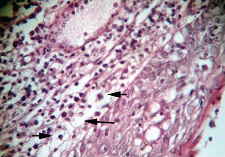
Photomicrograph of excised tissue showing atypical cells with vacuolated foamy cytoplasm suggestive of sebaceous differentiation (H and E stain; high magnification)

Photomicrograph of excised tissue showing atypical cells with vacuolated foamy cytoplasm suggestive of sebaceous differentiation (H and E stain; high magnification)