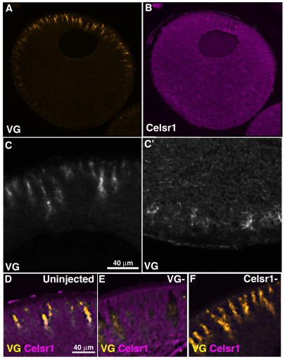
Fig. 1.
Vangl2 and Celsr1 distribution in wild-type, full grown (stage 6) Xenopus oocytes. (A,B) Histological sections of stage 6 oocytes showing immunostaining pattern for Vangl2 (VG) protein (A) and Celsr1 protein (B). (C,C′), Higher magnification of animal (C) and vegetal (C') hemisphere distribution of Vangl2 protein. (D-F), Images of animal hemisphere sections co-immunostained for Vangl2 and Celsr1 in uninjected control sibling (D), Vangl2-depleted (VG–; E) and Celsr1-depleted (Celsr1–; F) stage 6 oocytes.

