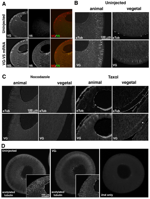
Fig. 2.
Vangl2 protein distributes with and maintains the acetylated microtubule cytoskeleton. (A) Co-immunostaining of sections from uninjected and Vangl2-V5 mRNA (20 pg)-injected stage 6 Xenopus oocytes using Vangl2 (VG) and V5 tag (V5) antibodies. (B) Co-immunostaining of sections of wild-type stage 6 oocytes for acetylated tubulin (aTub) and Vangl2 (VG) in the animal and vegetal hemispheres. (C) Co-immunostaining for acetylated tubulin and Vangl2 protein in sections of nocodazole-treated (5 μg/ml) and taxol-treated (2 μM/ml) stage 6 oocytes. (D) Optical sections (2.6 μm thickness) of stage 6 control and Vangl2-depleted (VG–) oocytes stained as whole mounts to show the acetylated tubulin cytoskeleton. Insets show higher magnification images of histological sections stained with the same antibody. Right-hand panel shows a control whole mount stained with secondary antibody only.

