Fig. 1.
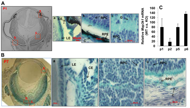
MAP3K1 expression in the developing eye. (A,B) Eyes isolated from the Map3k1+/ΔKD mice at (A) P1 and (B) P7 were subjected to whole mount X-gal staining and the histological sections were examined and photographed under a microscope. Arrows indicate the areas where the specific cell types are located. LE, lens epithelium; CE, ciliary epithelium; RPC, retinal progenitor cells; G, ganglion cells; RPE, retinal pigment epithelial cells; C, choroid. Scale bars: 20 μm. (C) RNA isolated from the retinas of wild-type and Map3k1ΔKD/ΔKD mice at different postnatal ages was subjected to real-time RT-PCR for Map3k1 expression. The levels of MAP3K1 in the knockout mice were similar to that in the control (no RNA) samples. In each age group, the expression of MAP3K1 in wild-type mice was compared with that in Map3k1ΔKD/ΔKD mice, set as 1. The results represent average of four samples at each genotype/age group+s.e.m.

