Fig. 2.
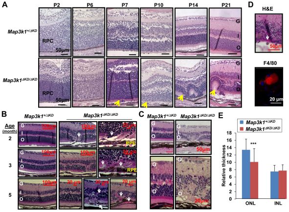
MAP3K1 is required for postnatal retina development. (A-D) The retinas of Map3k1+/ΔKD and Map3k1ΔKD/ΔKD mice at (A) P2-P21, (B) 2-5 months and (C,D) 1 year, were examined by Hematoxylin and Eosin staining and immunostaining, and were photographed under a microscope. RPC, retinal progenitor cells; G, ganglion cell layer; I, inner nuclear layer; O, outer nuclear layer; RPE, retinal pigment epithelium. Arrows indicate retinal lesions in the Map3k1ΔKD/ΔKD mice, including (A) malformed retina and rosette formation; and (B) photoreceptor dislocation, RPE breaks and blood vessel invasion. Different pictures of the Map3k1ΔKD/ΔKD retina are shown to illustrate various pathological features observed. The 1-year-old Map3k1ΔKD/ΔKD retinas have (C) clear thinner outer nuclear layer and retinal degeneration, and (D) F4/80-positive mononuclear macrophages (arrow, top panel, red, bottom panel). Cell nuclei are labeled with DAPI (blue). Scale bars: 50 μm (if not otherwise specified). (E) The thicknesses of the ONL and INL were measured in the 1-year-old Map3k1+/ΔKD and Map3k1ΔKD/ΔKD mice. Statistical analyses were carried out by comparing the average thickness between the genotypes. Data are mean+s.e.m. ***P<0.001.

