Fig. 3.
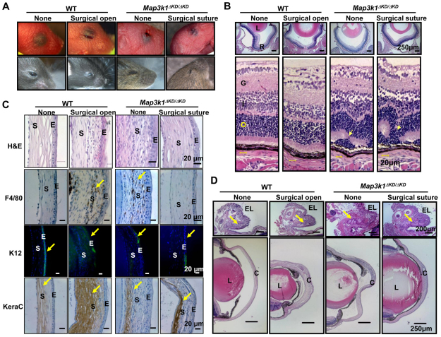
The role of a closed eyelid in postnatal eye development. Mice were subjected to surgical procedures to open the closed eyelid in wild type and suture the open eyelid in Map3k1ΔKD/ΔKD mice at P1 and suture was removed at P14. (A) Photographs of the eyes without or with the surgical procedures at P1 (top panels) and P15 (bottom panels). (B) The eyes at P15 were subjected to Hematoxylin and Eosin staining, and the retinas were examined under the microscope. L, lens; R, retina; G, ganglion layer; I, inner nuclear layer; O, outer nuclear layer. Clear retinal folding (arrows) is found in the Map3k1ΔKD/ΔKD mice regardless of whether the eyelid is sutured. (C) The corneas of the mice were subjected to Hematoxylin and Eosin staining, and immunohistochemistry with chromogenic reporters for macrophages (F4/80) and stromal keratocyte marker (keratocan), and with fluorescent reporter for corneal epithelial specific keratin 12 (K12, green) and DAPI for nuclei (blue). Surgical eyelid open in the wild-type mice causes severe corneal pathology (arrows), including macrophage invasion, loss of K12 expression and increased keratocan, similar to the knockout mice. S, corneal stroma; E, corneal epithelium. (D) The anterior surface of the eye was examined after Hematoxylin and Eosin staining. EL, eyelid; L, lens; C, cornea. MAP3K1 loss and eyelid suture do not affect Meibomian gland (arrows) formation in the eyelid (upper panels), but an opened eyelid at birth is clearly associated with corneal abnormalities (lower panels).

