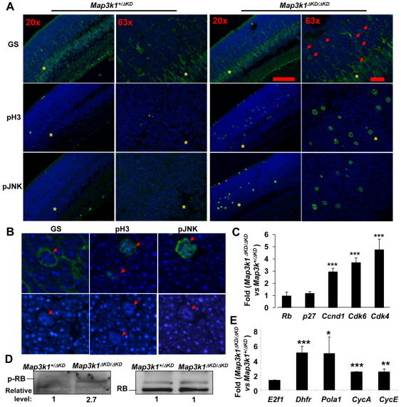
Fig. 8.
Activation of the E2F1 pathways in the retinas of Map3k1ΔKD/ΔKD mice. (A,B) Immunostaining of the P7 Map3k1+/ΔKD and Map3k1ΔKD/ΔKD retinas with various antibodies and pictures were taken under various magnifications, as indicated. The inner nuclear layer is labeled with asterisks and cells with condensed nuclei are labeled with arrows. (C,E) RNA isolated from the retinas of Map3k1+/ΔKD and Map3k1ΔKD/ΔKD mice at P7 were subjected to real-time RT-PCR to examine the expression of (C) genes that may affect RB, including Rb1, p27kip1, Ccnd1, Cdk4 and Cdk6, and (E) E2f1 and its target genes, Dhfr, Pola1, Ccna and Ccne. The levels of expression in the retinas of Map3k1ΔKD/ΔKD mice were compared with those in Map3k1+/ΔKD mice, which were set as 1. Results are average of at least four samples of each genotype. Data are mean+s.e.m. *P<0.05, **P<0.01, ***P<0.001. (D) Lysates of the P7 retinas were analyzed by western blotting for p-RB and total-RB, as indicated. The relative expression in the Map3k1ΔKD/ΔKD was compared with that in Map3k1+/ΔKD, which was set as 1.

