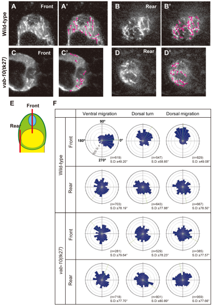
Fig. 5.
Direction of MT growth in DTCs. EBP-2-GFP comets were tracked and their directions were analyzed. (A-D′) Images representing 4 seconds of 200 mseconds/frame of EBP-2-GFP time-lapse sequences of the front (A,C) and rear (B,D) regions of wild-type (A,B) and vab-10(tk27) (C,D) DTCs that are migrating dorsally. `Front' is the nuclear side of the cell and `rear' is the opposite side. (E) Schematic presentation of the planes (shown in bars) of front and rear optical sections analyzed in A-D'. The direction of each shooting comet in A-D is indicated by arrows (A′-D′). (F) Directional distribution of comets in DTCs on the ventral muscle (ventral migration), at the dorsal turn and during dorsal migration is shown by Rose diagrams composed of 24 bins of 15° each. 0°, 90°, 180° and 270° correspond to distal, dorsal, proximal and ventral directions, respectively. The concentric circles were drawn with 5% increments between them. Green lines represent the average angles.

