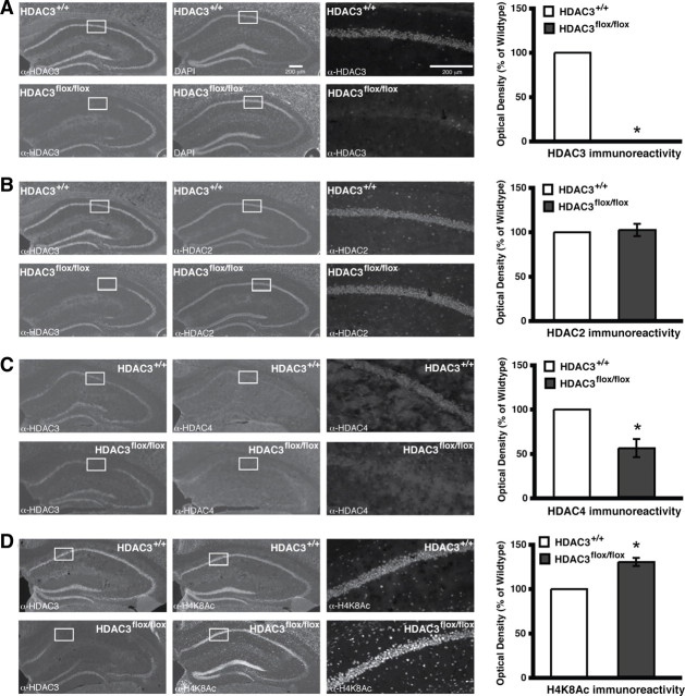
Figure 1.
Intrahippocampal AAV2/1–Cre infusion in HDAC3flox/flox mice results in a complete, focal deletion of HDAC3 that correlates with increased histone acetylation. Images are 4×, except the right panels, which are 20× magnifications of the regions boxed in white. Histograms depict quantification of optical density as a percentage of wild type. A, Representative images showing DAPI labeling and HDAC3 immunoreactivity in hippocampi of AAV2/1–Cre infused HDAC3+/+ and HDAC3flox/flox mice. HDAC3 labeling is found throughout CA1, CA3, and the dentate gyrus, and no immunoreactivity is found in the AAV2/1–Cre infusion site of HDAC3flox/flox mice. *p < 0.05. B, Representative images showing HDAC2 immunoreactivity in hippocampus is unchanged in AAV2/1–Cre-infused HDAC3flox/flox mice. C, However, HDAC4 immunoreactivity is decreased in the region of the HDAC3 deletion. *p < 0.05. D, Furthermore, acetylation at H4K8 is increased specifically in the AAV2/1–Cre infusion site of HDAC3flox/flox mice. *p < 0.05.

