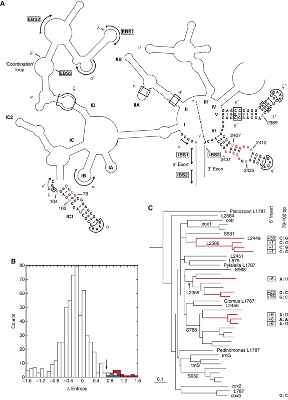
Figure 1.
Identification of a candidate site for binding the branchpoint-carrying domain of a group II intron. (A) Schematic secondary structure of the Pl.L1787 (Pl.LSU/2) ribozyme, a representative mitochondrial member of subgroup IIB1. Only the sequences of domains V and VI and the distal part of subdomain IC1 are shown, the asterisk next to domain VI indicates the branchpoint. Greek letters and arrows correspond to prominent tertiary interactions, which are generally conserved in group II introns (Michel et al, 2009). Sites in red and orange are those at which the difference in sequence entropy between the set of introns with and without a 5′ terminal insert exceeds 1.0 or is included in the 0.70–1.0 range, respectively (see (B)). (B) Statistical distribution over aligned ribozyme sites of the difference in sequence entropy between sets of introns with and without a 5′ terminal insert. Ordinates: number of sites; abscissa: difference in sequence entropy at homologous sites between the two intron sets, calculated as in Materials and methods (numbers are positives when site entropy is larger for the set of introns with a 5′ insert). The arrow points to the 0.70 differential entropy threshold (for sites highlighted in (A); red and blue rectangles correspond to sites in domains VI and IC1, respectively). (C) Phylogenetic relationships of mitochondrial subgroup IIB1 introns based on an alignment of their ribozyme sequences (the tree is redrawn from Li et al, 2011). Introns and intron clades are designated by their host gene (Li et al, 2011). Thick red lines correspond to lineages of introns that possess a 5′ terminal insert, the length of which is indicated at right (boxed numbers). When not G and U, the nucleotides at positions 79 and 100 (of the Pl.L1787 ribozyme) are indicated at the far right.

