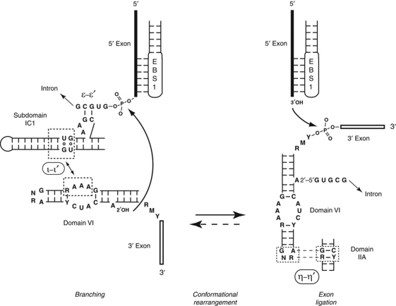
Figure 5.
Conformational rearrangements and tertiary interactions involving domain VI. Tentative delimitation of the ι and ι′ motifs is based on our modelling of the interaction in Figure 3A. During the splicing process, domain VI is successively bound by ribozyme subdomain IC1 (ι–ι′ interaction—this work—which positions domain VI for the branching step) and subdomain IIA (η–η′ interaction—Chanfreau and Jacquier, 1996—which positions domain VI for exon ligation; a 90° rotation was chosen for convenience of drawing, the actual value must be less, see Figure 3A). In reverse splicing into a DNA or (possibly) RNA target, formation of ι–ι′ should follow that of η–η′ (dashed arrow). Bases shown are consensus ones for mitochondrial subgroup IIB1 introns (Li et al, 2011). Curved arrows symbolize reactions.

