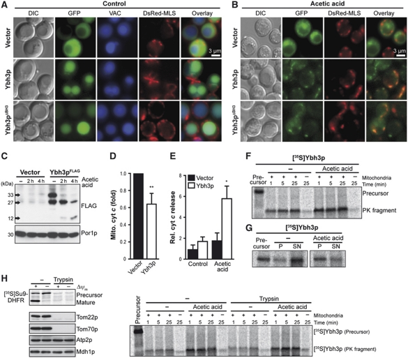
Figure 4.
Ybh3p translocates to mitochondria and is imported in a TOM-receptor-independent way. (A, B) Fluorescence microscopy of yeast cells expressing GFP-tagged Ybh3p or Ybh3pΔBH3 or harbouring the vector control for 16 h before (A) and after (B) treatment with acetic acid. Vacuoles were counterstained using Celltracker Blue. For visualization of mitochondria, the mitochondrial marker DsRed Su1-69 (DsRed-MLS) was co-expressed. (C) Immunoblot analysis of mitochondrial fractions harvested from cells overexpressing FLAG-tagged Ybh3p without or with acetic acid treatment for 2 and 4 h before subcellular fractionation. (D) The cyt c content of mitochondrial fractions harvested from cells overexpressing Ybh3p compared with isogenic vector control after 4 h of acetic acid treatment determined by immunoblot analysis and subsequent quantification of cyt c signal compared with porin signal (Por1p, mitochondrial marker) (mean±s.e.m., n=6). (E) The cyt c content in supernatants after release of cytosolic content via short permeabilization. Cells overexpressing Ybh3p or harbouring the empty vector were pre-treated or not with 120 mM acetic acid for 2 h prior to permeabilization followed by immunoblot analysis. Cyt c signal in supernatants was normalized to cyt c content in whole cell extracts (mean±s.e.m., n=5). (F, G) 35S-labelled Ybh3p was incubated with isolated yeast mitochondria, followed by treatment with proteinase K (F) or carbonate extraction (G) and analysis by NuPAGE and autoradiography. For carbonate extraction, supernatants (SN) and pellets (P) were analysed. Cells were grown in the presence or absence of acetic acid prior to isolation of mitochondria. (H) Yeast mitochondria isolated from cells grown in the presence or absence of 90 mM acetic acid were incubated with 35S-labelled Su9-DHFR for 30 min or with 35S-labelled Ybh3p for the indicated time followed by treatment with proteinase K and analysis by NuPAGE and autoradiography. Isolated mitochondria were incubated with or without trypsin prior to the import reaction. In addition, mitochondria were analysed for Tom22p, Tom70p, Atp2p and Mdh1 using immunoblotting. Where indicated the membrane potential (Δψ) was dissipated prior to the import reaction. See also Supplementary Figure S4. *P<0.05 and **P<0.01.

