Figure 5.
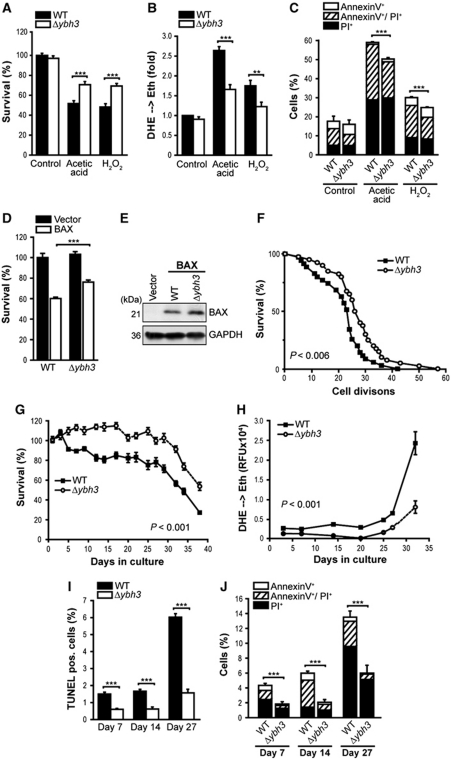
Deletion of YBH3 protects against H2O2, acetic acid, BAX expression and ageing. (A, B) Survival determined by clonogenicity (A) and quantification of ROS accumulation (DHE → Eth) (B) of wild-type (WT) and Δybh3 cells treated or not with acetic acid or H2O2 (mean±s.e.m., n=8). (C) Flow cytometric quantification of phosphatidylserine externalization and loss of membrane integrity using AnnexinV/PI co-staining of cells described in (A) (mean±s.e.m., n=4). (D) Survival determined by clonogenicity upon heterologous expression of murine BAX in WT and Δybh3 cells. Viability was determined after 20 h of BAX expression (mean±s.e.m., n=8). (E) Immunoblot analysis of WT and Δybh3 cells expressing BAX. (F) Replicative lifespan of WT and Δybh3 cells. A representative experiment is shown with n⩾40. Indicated P-value (calculated using Tarone-Ware) refers to median lifespan of Δybh3 compared with WT. (G, H) Survival determined by clonogenicity (G) and quantification of ROS accumulation using DHE → Eth conversion (H) of WT and Δybh3 cells during chronological ageing on glycerol-containing media. Colony forming units of WT cells at day 1 were set to 100%. One representative ageing experiment is shown, with data representing mean±s.e.m. of six independent cultures. (I, J) Flow cytometric quantification of TUNEL staining (I) and AnnexinV/PI-co-staining (J) of chronologically aged WT and Δybh3 cells at indicated days during ageing (mean±s.e.m., n=4). See also Supplementary Figure S5. **P<0.01 and ***P<0.001.

