Figure 1.
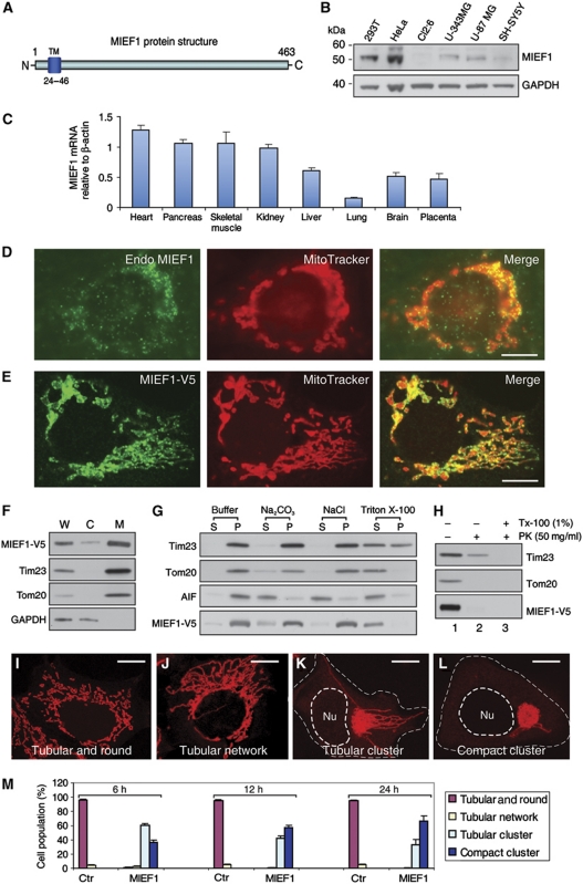
The protein structure, expression and subcellular localization of MIEF1. (A) MIEF1 is an integral outer mitochondrial membrane protein with an N-terminal TM domain. (B) Endogenous MIEF1 in various cell lines was immunoblotted using anti-MIEF1 antibody. (C) Real-time PCR analysis of MIEF1 expression in normal human adult tissues. Levels of MIEF1 mRNA were determined relative to β-actin. Data were from three independent experiments. (D, E) Both endogenous MIEF1 and exogenous MIEF1-V5 were localized to mitochondria, stained with anti-MIEF1 antibody (green) and MitoTracker Red (red). (F) The distribution of MIEF1-V5, Tim23, Tom20 and GAPDH was analysed in whole-cell lysate (W), cytosolic fraction (C) and mitochondrial fraction (M). (G) Mitochondrial fractions prepared from 293T cells expressing MIEF1-V5 were resuspended in the mitochondrial buffer (buffer) alone as control, or in buffers containing 0.1 M Na2CO3 (pH 11.5), 1 M NaCl or 1% Triton X-100 followed by centrifugation, and the membrane pellets (P) and supernatant fractions (S) were immunoblotted with indicated antibodies. (H) Mitochondrial fractions were digested with PK in the absence (lane 2) or presence (lane 3) of 1% Triton X-100 (Tx-100) for 30 min or with mock control (lane 1) and analysed for MIEF1-V5, Tim23 and Tom20. (I–L) Mitochondrial morphology in 293T cells transfected with empty vector (I, J) and MIEF1-V5 (K, L) was analysed by confocal microscopy after double staining with MitoTracker and anti-V5 antibody (not shown). Outlines of the nucleus (Nu) and the cell are drawn in dash line. (M) Percentages (mean±s.e.m.) of 293T cells with indicated mitochondrial morphologies at indicated time points post-transfection for empty vector (Ctr) transfected cells (n=495 for 6 h; n=558 for 12 h; n=568 for 24 h) and for MIEF1-V5 transfected cells (n=427 for 6 h; n=735 for 12 h; n=776 for 24 h). Data were from three independent experiments. Bars, 10 μm.

