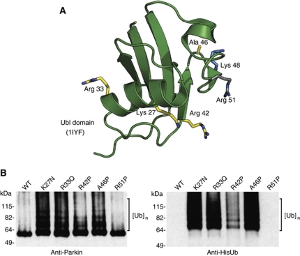
Figure 2.
Pathogenic mutations in the Ubl domain relieve autoinhibition. (A) NMR structure of human Parkin showing the location of each of the pathogenic mutations in yellow, Lys48 in blue, and the site of the silent mutation in grey (PDB code 1iyf (Sakata et al, 2003)). (B) Western blot analysis of Parkin autoubiquitination shows that pathogenic point mutations render Parkin active for autoubiquitination while R51P does not. Ubiquitin conjugates are detected using Parkin and His antibodies and are indicated by brackets. Ube2L3 is the E2 in the assay.

