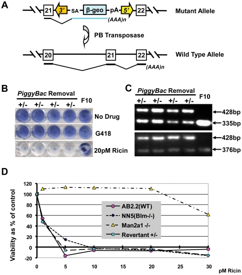
Figure 3. PBase reversion of mannosidase 2, alpha 1 mutation.
A, illustration of the allele bearing the Man2α1PiggyBac insertion before and after reversion with PBase. B, methylene blue staining of four revertant heterozygous clones, and the original ricin resistant F10 clone, either before or after G418 or 20 pM ricin exposure. C, triple primer PCR demonstrating heterozygosity after reversion. D, viability as percentage of control calculated from neutral red staining of AB2.2, NN5, F10 Man2α1 homozygous mutant and revertant cells after exposure to different concentrations of ricin (0 or 1–30 pM).

