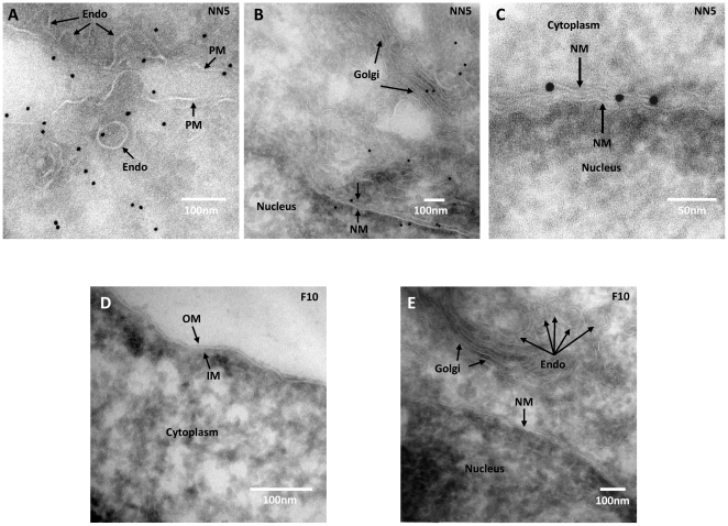
Figure 6. Anti-ricin immunogold staining of either NN5 or ricin resistant F10 cells.
A–C, images showing ricin particles located in various sites within NN5 cells treated with 20 pM ricin for 1 h at 37°C. D and E ricin-resistant F10 cells exposed to 20 pM ricin and processed for anti-ricin staining at the same time. NM, PM, Endo, OM and IM represent nuclear membrane, plasma membrane, endosome, outer membrane and inner membrane respectively (small arrows). The nucleus, cytoplasm and Golgi are also indicated.

