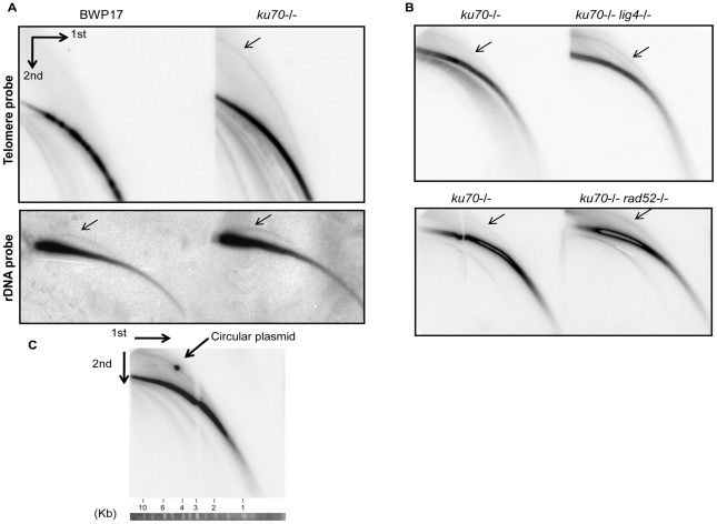
Figure 6. Identification of t-circles in the ku70 null mutants.
(A) (Top) Genomic DNA samples were prepared from the parental wild type strain (BWP17) and its ku70-/- derivative (LCF2.1), digested with AluI and NlaIII, and then subjected to 2-D electrophoresis followed by Southern blotting to identify linear and circular telomeric DNA. (Bottom) The sample DNA samples were subjected to 2-D gel electrophoresis (without prior digestion) and analyzed using an rDNA probe. (B) Genomic DNA samples from the indicated strains were subjected to 2-D gel electrophoresis followed by Southern blotting to assess the levels of linear and circular telomeric DNA. All mutant strains were derived from the CAI4 parental strain, as follows: ku70-/- (LCD2A.1); ku70-/- lig4-/- (CEA2.5); ku70-/- rad52-/- (JLT2.1). (C) Genomic DNAs from the ku70-/- strain (LCD2A.1) were digested and analyzed by 2-D gel electrophoresis together with a nicked 4.4 kb circular plasmid (pGEM-URA3, isolated by gel electrophoresis). Radioactive probes for the plasmid and for telomere DNAs were both included in the hybridization mixture. A strip of DNA size standards corresponding to the first round of electrophoresis is shown at the bottom.

