Fig. 2.
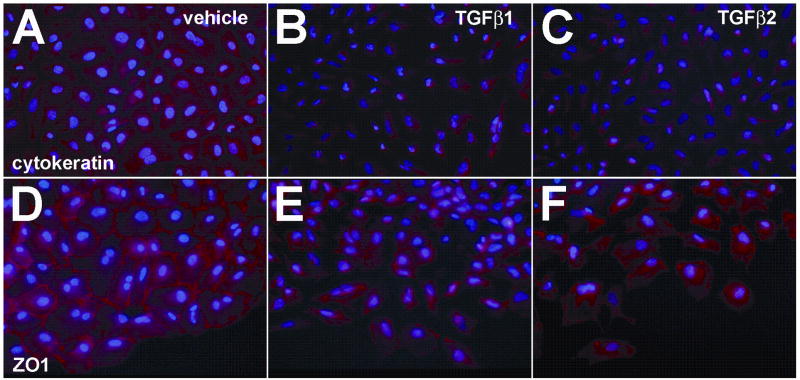
TGFβ alters cytokeratin and ZO1 expression in PE cells. PEs were incubated with vehicle, TGFβ1 or TGFβ2 for 24 hours. After fixation, explants were processed to detect either cytokeratin or ZO1 expression by immunofluorescence. A, Epithelial cells in PEs incubated with vehicle express cytokeratin abundantly. B,C, Cells from explants incubated with 200 pM TGFβ1 (B) or TGFβ2 (C) separate from the epithelial sheet, become spindle shaped, and have decreased expression of cytokeratin. D, ZO1 immunoreactivity in vehicle incubated explants is located at the periphery of cells demarcating cell-cell contact points between adjacent epithelial cells. E,F, Cells from explants incubated with 200 pM TGFβ1 (E) or TGFβ2 (F) separated from the epithelial sheet and changed shape. ZO1 immunoreactivity was less prominent at the cell periphery, consistent with a loss of cell-cell contacts and the transition from an epithelial to a mesenchymal cell phenotype. All panels, 400x.

