
Figure 1.
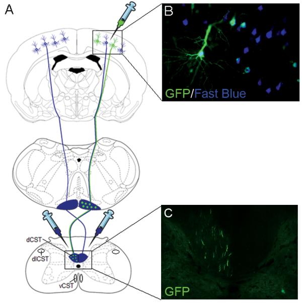
Overview of the experiment. (A) Schematic showing the rodent CST originating from the pyramidal CSNs in layer V of the sensorimotor cortex, decussating at the spinomedullary junction, forming the main dCST and the dlCST and vCST minor components. Rats received six unilateral viral vector injections into the sensorimotor cortex to transduce the CSNs and bilateral C1/C2 intraspinal injections of the retrograde tracer Fast Blue to label the CSNs. (B) Image showing transduced, retrogradely labelled CSNs. (C) Image showing GFP-positive CST fibres in the contralateral dCST of the cervical spinal cord.