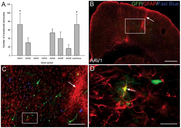
Figure 7.
Viral vectors did not exhibit a strong astrocytic tropism. (A) Quantification of transduced astrocytes in the injected cortical hemisphere. GFP and GFAP positive cells were counted and the mean number of transduced astrocytes plotted for each viral vector. Values represent mean and SEM, analysis was performed using one way ANOVA with Tukey post-hoc tests * P < 0.05, n = 3/group. (B) A GFP and GFAP stained cortical hemisphere from an AAV1 transduced rat. Enhanced GFAP staining can be observed along the needle track (arrow). Scale bar: 500 μm. (C) Higher-magnification image of box in (B) showing the viral injection site and the enhanced GFAP staining around the needle track (arrow). Scale bar: 150 μm. (D) Higher-magnification image of box in (C) showing a transduced, GFAP positive astrocyte (arrow). Scale bar: 25 μm.

