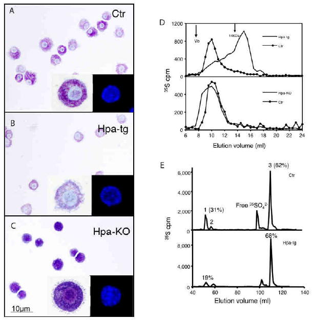
Figure 1. Characterization of FSMCs.

In vitro differentiated FSMCs from Ctr (A) hpa-tg (B), and hpa-KO (C) embryos were stained with toluidine blue. Magnification 40×. Inserts represent enlarged or DAPI-stained cells. (D) Gel chromatography analysis of metabolically 35S-labeled heparin from FSMCs. Upper panel: Ctr vs. hpa-tg; lower panel: Ctr vs. hpa-KO. (E) Analysis of disaccharides by anion-exchange HPLC. The numbered peaks represent: 1. -GlcA-GlcNS6S-; 2. -IdoA-GlcNS6S-; 3. -IdoA2S-GlcNS6S- sequences of the intact heparin chains. The percentages of the respective components in total recovered disaccharides are indicated for peaks 1 and 3. Upper panel: Ctr; lower panel: hpa-tg.
