Figure 3.

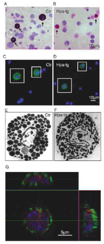
Morphological examination of peritoneal MCs.
Peritoneal cells were stained with May Grünwald/Giemsa: (A), Ctr; (B), hpa-tg (arrows point to MCs); or with FITC-conjugated anti-c-kit antibody: (C), Ctr; (D), hpa-tg; MCs are illustrated by squares. Original magnification 40×. (E, F) TEM analysis of Ctr (E) and hpa-tg (F) peritoneal MCs. Original magnification 3000×. (G) Hpa-tg MCs double immunostained for c-kit (green) and heparanase (red) examined by confocal microscopy (z-scan). The nuclei were stained with DAPI (blue).
