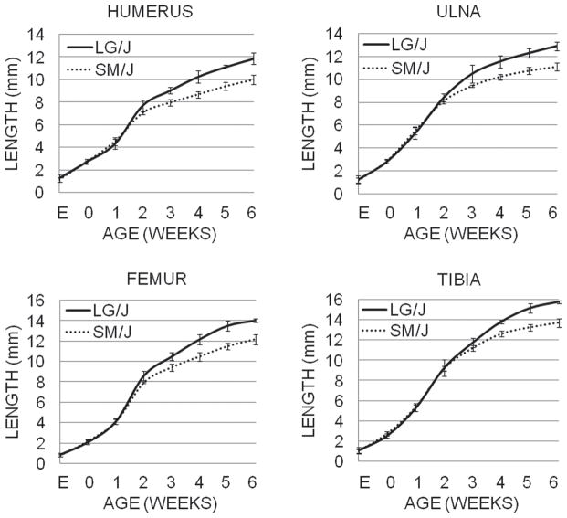
Figure 3.
The average lengths (in mm) of the humerus, ulna, femur, and tibia at various ages are shown for LG/J (solid line) and SM/J (dotted line) animals. Males and females are represented approximately equally for each age in the cohort (cohort sizes listed in Supplemental Table 1), with 3 extra males included in the 3–5 week age categories. E refers to 17.5 days post conception, and 0 refers to the day of birth; all other ages are given in weeks. Day E17.5-1 week lengths are diaphysis lengths, while 2–6 week lengths are epiphysis-to-epiphysis lengths. Standard deviations are shown by vertical bars and are listed in Supplemental Table 1.

