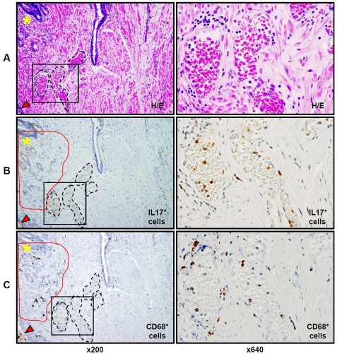
Figure 5.
H&E and IHC staining for IL-17 and CD68 in lumens of capillaries in formalin-fixed paraffin-embedded whole mount radical prostatectomy specimens from patients with prostate cancer. A - H&E staining, B - staining for IL-17 (light brown color) and C - staining for CD68 (dark brown color) expressing cells in lumens of microvessels (black dotted circles, ×200) located in benign tissue between the areas of prostate adenocarcinoma (not visible at right side of the slide), proliferative inflammatory atrophy (PIA) lesion (red arrowheads, ×200) and MNC hot spot (yellow asterisks, ×200). Stromal CD68+ tissue macrophages (dark brown color in red circles, ×200) adjacent to the MNC hot spot show no co-expression of IL-17 protein. Left column presents H&E staining and immunostainingfor IL-17 and CD68 in prostate tissue with magnification ×200. Right column depicts H&E staining and immunostainingfor IL-17 and CD68 in prostate tissue located inside of black squares of the left columns with magnification ×640. Details are described in “Materials and Methods” section.

