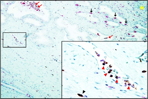
Figure 6.
IHC staining for IL-17 and CD68 inside microvessel at the PIA lesion of formalin-fixed paraffin-embedded whole mount radical prostatectomy specimens from patients with prostate cancer (magnification ×125). IL-17 (brown color) and CD68 (pink color) proteins are co-expressed (dark brown or black colors) in monocyte/macrophages inside capillaries located in stroma adjacent to PIA lesion. Red arrows indicate intra-glandular IL-17 expressing CD68+ macrophages. Black arrows indicate intra-epithelial IL-17-non-producing CD68+ macrophages. Yellow asterisk indicates site of accumulation of mononuclear cells (“hot spot”) in the prostate stroma. Insertion represents microvessel located inside of the black square: red arrowheads indicate IL-17 producing CD68+ monocyte/macrophages inside of the capillary space; black arrowhead indicates stromal IL-17 expressing CD68+ macrophage (magnification ×800). The figure shows that IL-17 expressing cells can be readily detected inside lumens of capillaries located in the peripheral zone of the prostate, and that CD68 monocytes/macrophages inside the stromal capillaries near PIA lesion express IL-17 protein. Details are described in “Materials and Methods” section.

