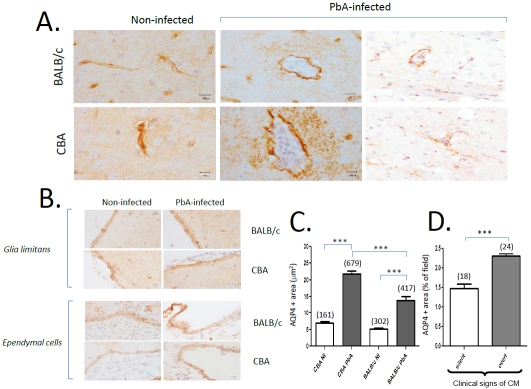
Figure 2.
Patterns of AQP4 expression in CM-S and CM-R mice upon PbA infection. A. Distinct patterns of AQP4 expression: selectively increased expression in perivascular spaces in CBA mice with CM, on day 7 p.i. The AQP4 expression in astrocytic foot processes is higher in CBA than in BALB/c mice. AQP4 is revealed in brown (DAB) except on the two right panels, where it is revealed in red (LPR) and GFAP in brown (DAB), representative of 5 mice. B. AQP4 patterns in glia limitans and ependymal cells. DAB staining. C. Quantitation of AQP4 expression by planimetry in noninfected (NI) versus PbA-infected mice. The incubation conditions were identical between all sections and all animals by incubating simultaneously for all immunostainings. Numbers in parentheses indicate the numbers of vessels studied. *: p<0.01,***: p < 0.0001. D. AQP4 expression in relation to clinical expression of CM. CBA mice, 7 days p.i., with clinically silent versus clinically overt neurological syndrome of CM. Numbers in parentheses indicate the numbers of vessels studied. Kruskall-Wallis test, non-parametric one-way ANOVA with Bonferroni test.

