Figure 2.
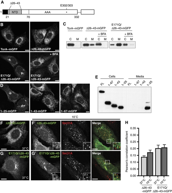
Residues 26–43 of torsinA's NTD are necessary and sufficient for ER retention. (A) Schematic view of torsinA sequence: signal peptide (1–20, black), NTD (21–43, grey), linker region (44–70), AAA domain (71–332), ΔE mutation (*, 302/303), and C-terminal mGFP tag. (B) Confocal microscopy of torsinA–mGFP, Δ26–43-torsinA–mGFP, E171Q/Δ26–43-torsinA–mGFP, and E171Q/Δ26–43-torsinA–mGFP in the presence of BFA. (C) Immunoblot of the indicated GFP fusion proteins in cell lysates or media immunoprecipitates. (D) Confocal microscopy of torsinA's signal sequence (1–25), NTD (1–43), or NTD plus linker region (1–67) fused to mGFP. Scale bars, 10 μm. (E) Immunoblot of the indicated GFP fusion proteins in cell lysates or media immunoprecipitates. (F,G) Confocal microscopy of cells expressing Δ26–43-torsinA–mGFP or E171Q/Δ26–43-torsinA–mGFP at 37°C. (F′,G′) Costaining with Sec31A after 2 h incubation at 10°C. Scale bars, 10 μm. (H) Quantification of colocalization of the indicated GFP-tagged proteins with Sec31A. N>20 cells for each condition. Bars indicate standard error of the mean.

