Figure 4.
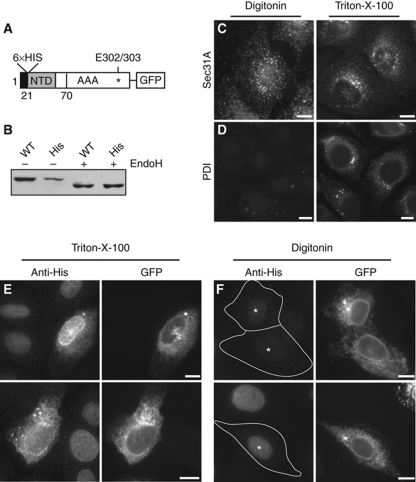
TorsinA's N terminus is not accessible from the cytosol and is therefore monotopically associated with the ER bilayer. (A) Schematic diagram of His–torsinA–mGFP construct. (B) His–torsinA–mGFP migrates at the same apparent molecular weight as wild-type torsinA–mGFP and is similarly sensitive to EndoH. (C, D) Selective permeabilization with digitonin allows detection of the cytosolic Sec31A epitope (C) but not the lumenal PDI epitope (D). (E, F) Cells expressing His–torsinA–mGFP were treated with Triton-X-100 (E) or with digitonin under conditions that permeabilize only the plasma membrane (F). The N-terminal His tag was detected using anti-His antibody and gave specific signal in Triton-X-100 but not in digitonin-treated samples. Transfected cells are indicated with asterisks (*); note that background nuclear staining by the anti-His antibody is unrelated to His–TorA–GFP expression. Scale bars, 10 μm.

