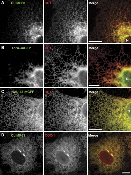
Figure 8.
Preference of torsinA and COX-1 for ER sheets. (A–C) Confocal microscopy of COS-7 cells stained for or expressing the indicated GFP-tagged proteins, zoomed in to show a section of the perinuclear and peripheral ER. ‘N’ indicates the nucleus. (D) Confocal microscopy of COS-7 cells expressing untagged COX-1, costained for CLIMP-63. Scale bars, 10 μm.

