
Figure 3.
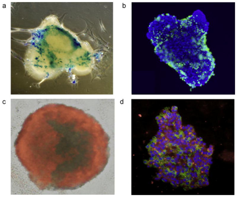
Microscopic appearance of labeled islets. Prussian Blue staining (a, blue = ferric iron in iron oxide) and anti-dextran immunostaining (b, green = dextran coat of Feridex®, blue = DAPI nuclear counterstain) of Feridex®-labeled human islets reveal scattered inhomogeneous staining of cells. (c) When labeled with PFPE/rhodamine, all cells exhibit uptake and positive staining of label. (d) Immunostaining of PFOB/rhodamine (red)-labeled islet cells demonstrates functional insulin production (green, with DAPI blue nuclear counterstain).