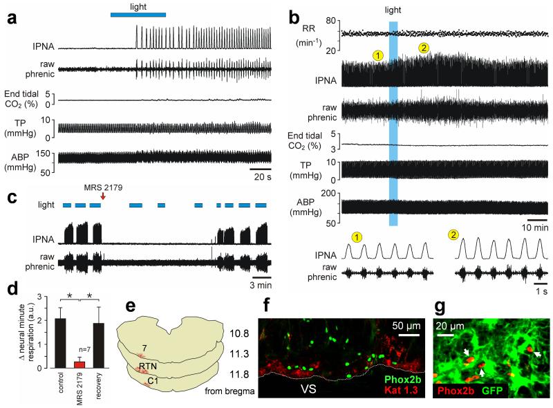
Fig. 4. Optogenetic activation of VS astrocytes stimulates breathing in vivo.
(a) Unilateral photostimulation of VS astrocytes expressing ChR2(H134R)-Katushka1.3 is sufficient to trigger respiratory activity from hypocapnic apnea in an anesthetized rat. Hypocapnic apnea was induced by mechanical hyperventilation to reduce arterial levels of PCO2/[H+] below the apneic threshold. IPNA – integrated phrenic nerve activity. TP – tracheal pressure. ABP – arterial blood pressure. (b) Lasting effect of light activation of VS astrocytes in an animal breathing normally. RR – respiratory rate. (c) Time-condensed record illustrating effects of repeated stimulations of VS astrocytes on phrenic nerve activity before and after a single application of MRS2179 (100 μM, 20 μl) on the VS. Note spontaneous recovery of the response over time. (d) Summary data of MRS2179 effect on the increases in neural minute respiration (the product of phrenic frequency and amplitude) evoked by light-activation of VS astrocytes (*p < 0.05). (e) Rostro-caudal distribution of astrocytes expressing ChR2(H134R)-Katushka1.3 in the brainstem of the rat from the experiment shown in (a). 7 – facial nucleus. RTN – retrotrapezoid nucleus. C1 - catecholaminergic cell group. (f) ChR2(H134R)-Katushka1.3 (Kat 1.3) expression in astrocytes is identified by red fluorescence distributed near the VS in close association with Phox2b-immunoreactive neurons (green nuclei). Coronal brainstem section. (g) Phox2b-expressing chemoreceptor RTN neurons (red nuclei) embedded in the astrocytic network (astrocytes are transduced with Case12 in this example to reveal their morphology).

