
Figure 3.
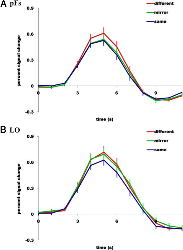
Hemodynamic time courses (percentage signal change) of two object-selective regions of cortex, pFs (A) and LO (B) to (1) two completely different images of objects (red line labeled “different”), (2) the same image of an object presented twice (blue line labeled “same”), and (3) an object followed by the mirror-reversed version of the same object (green line labeled “mirror”). Note tolerance to mirror-image reversals in pFs, yet sensitivity to mirror-image reversals in LO.