
Fig. 4.
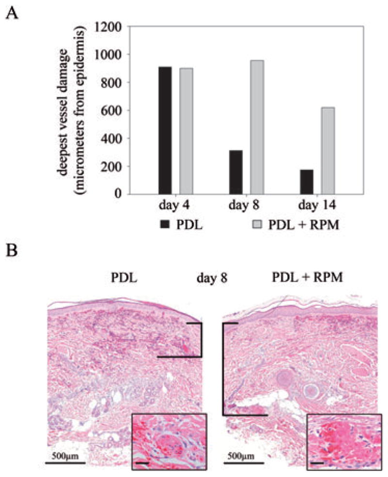
Topical RPM increases PDL-induced endothelial damage. Skin was exposed to PDL alone (8 J/cm2 energy density) or to a combination of PDL (8 J/cm2 energy density) and topical RPM. Punch biopsies of exposed skin areas were taken after 4, 8 and 14 days. A) Deepest measurable vascular damage in both treatment groups displayed in bar graph format, in micrometer from the base of the epidermis. Results 4, 8 and 14 days post-PDL-exposures are shown. B) Representative H&E stained sections from each treatment protocol are shown 8 days post-PDL. The vertical bars indicate the distance from the base of the epidermis to the deepest damaged vessel (inset shows high power view of destroyed vessels). Bar: main image: 500 μm; inset: 100 μm.