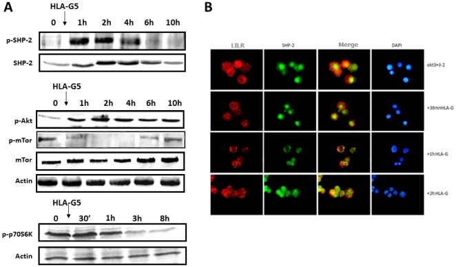
Figure 4. A: Time-course modification of pSHP-2, p-Akt, p-mTOR, and p70S6K on activated T cells incubated with HLA-G5-coated beads.
T lymphocytes were activated before incubation with OKT3/IL-2 for 72 h. In the presence of HLA-G, p-mTOR and p70S6K were transiently dephosphorylated, but Akt was not. This result correlates with SHP-2 phosphorylation. B: Intracellular distribution of ILT-2 and SHP-2 in the presence of HLA-G as assessed by immunofluorescence. In stimulated T cells incubated with HLA-G5-coated beads, SHP-2 was redistributed intracellularly and translocated from the nucleus to the cytoplasm, whereas it was only partially redistributed after 1 h with LILRB1.

