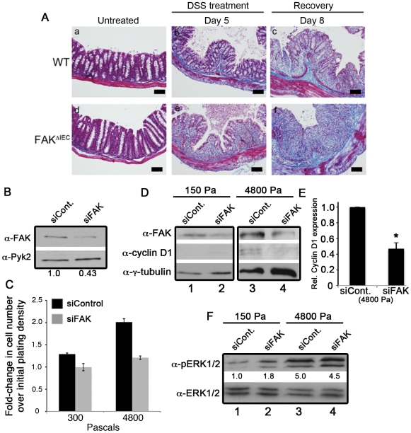
Figure 6. Increased tissue rigidity leads to FAK-dependent cell proliferation.
(A) Colon sections from untreated and DSS-treated WT and FAKΔIEC mice were stained with Masson's trichrome stain. Collagen appears blue, muscle stains dark red, cytoplasm stains pink and nuclei appear dark brown. Bars represent 50 µm. (B) Caco-2 cells transfected with siControl or siRNA targeting FAK (siFAK) were lysed 72 hours post-siRNA transfection and immunoblotted for total FAK and Pyk2. (C) 24 hours post-transfection, cells were inoculated (6 wells per conditions) onto a soft-plate96, incubated for 48 hours, and quantified using the CyQuant NF proliferation assay. Data are representative of 2 independent experiments. (D) Caco-2 cells were transfected with siControl or siFAK for 24 hours before plating onto polyacrylamide gels with rigidities of 150 Pa or 4800 Pa, and cultured for a further 48 hours. Cells were then lysed and immunoblotted for FAK, cyclin D1 and tubulin. (E) Cyclin D1 levels were normalized to the amount of total tubulin and expressed relative to the amount of cyclin D1 in siControl-treated cells Data are representative of 3 independent experiments. Asterisks denote values that are significantly different from siControl-treated cells (P<0.05). (F) Caco-2 cells were transfected and plated as described in part D. Cells were then lysed and immunoblotted for phopho- and total ERK1/2. Phospho-ERK1/2 levels were normalized to total ERK1/2 and expressed relative to the amount of phospho-ERK1/2 in siControl-treated cells plated onto the 150 Pa substrate (see numbers under the immunoblot).

