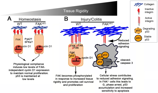
Figure 7. Increased tissue rigidity drives cell cycle progression in a FAK-cyclin D1-dependent manner.
(A) Under homeostatic or physiological conditions, where tissue compliance is relatively high, FAK and integrin receptors are minimally associated and cyclin D1/proliferation are kept to basal levels. p53 levels are also maintained at low levels. (B) Induction of colitis leads to the deposition of collagen and other ECM components. The resultant increased tissue rigidity promotes FAK-integrin complexes, which in turn induces FAK phosphorylation and promotes cell survival. FAK auto-phosphorylation can result in Akt activation and subsequent phosphorylation of Mdm2. FAK translocation to the nucleus also allows FAK to function as a scaffold, stabilizing p53-Mdm2 complexes. Both of these FAK signaling pathways enhance cell survival by keeping p53 levels low. Finally, FAK contributes to the induction of cyclin D1 expression by upregulating transcription factors such as krupple-like factor 8 (KLF-8). In the absence of FAK, the failure to increase cyclin D1 levels significantly attenuates proliferation and impairs the healing response.

