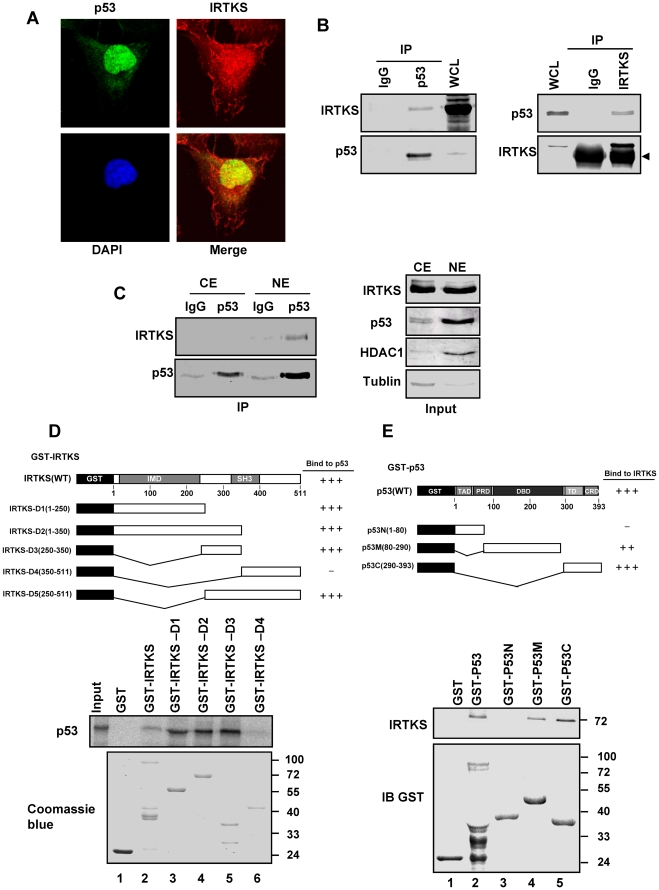
Figure 2. IRTKS directly interacted with p53.
(A) Co-localization of IRTKS and p53. HT1080 cells were immunostained with anti-p53 antibody and anti-IRTKS antibody followed by Alexa-488 conjugated anti-mouse antibody and Alexa-546 conjugated anti-rabbit antibody. Images were taken through a confocal microscope. (B) Association of endogenous ITRKS and endogenous p53 in HEK 293 cells. Whole cell lysates (WCL) and immunoprecipitations (IP) with control IgG, p53 antibody, preimmune serum or IRTKS antibody were analyzed by Western blotting. The arrowhead indicates IgG heavy chain. (C) IRTKS interacted with p53 in nucleus. Immunoprecipitations of the nuclear and cytoplasmic extracts from HT1080 cells with p53 antibody were analyzed with IRTKS antibody by Western blotting. (D) Direct interaction between p53 and IRTKS revealed by GST-pulldown assays. Full-length or truncated GST-IRTKS proteins were incubated with in vitro translated 35S-labelled p53 in NETN buffer, and then precipitated with glutathione-Sepharose beads. Bound proteins were resolved by 10% SDS-PAGE followed by autoradiography. GST-IRTKS proteins were visualized by Coomassie blue staining. (E) Interaction site mapping on p53. GST-pulldown assays were performed with recombinant His-IRTKS and full-length or truncated GST-p53. The pulldown samples were subjected to Western blotting with anti-IRTKS and anti-GST antibodies.

