Figure 2.
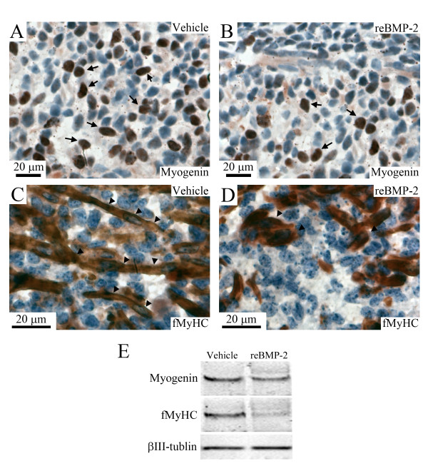
Recombinant BMP-2 suppressed the expressions of myogenin and fMyHC in cultured tongue. Immunostaining image for myogenin (A, B) and fMyHC (C, D) in the middle portion of E13 tongues cultured for 8 days in BGJb medium containing the vehicle (A, C) and 4 μg/ml of recombinant BMP-2 (B, D). Brown color indicates immunostaining image for myogenin and fMyHC. Arrows in A and B indicate myogenin-positive nuclei and arrowheads in C and D indicate elongated myotubes and myofibers. Western blotting pattern of myogenin, fMyHC, and βIII-tubulin in the whole portion of E13 tongues cultured for 8 days in BGJb medium containing the vehicle and 4 μg/ml of recombinant BMP-2 (E).

