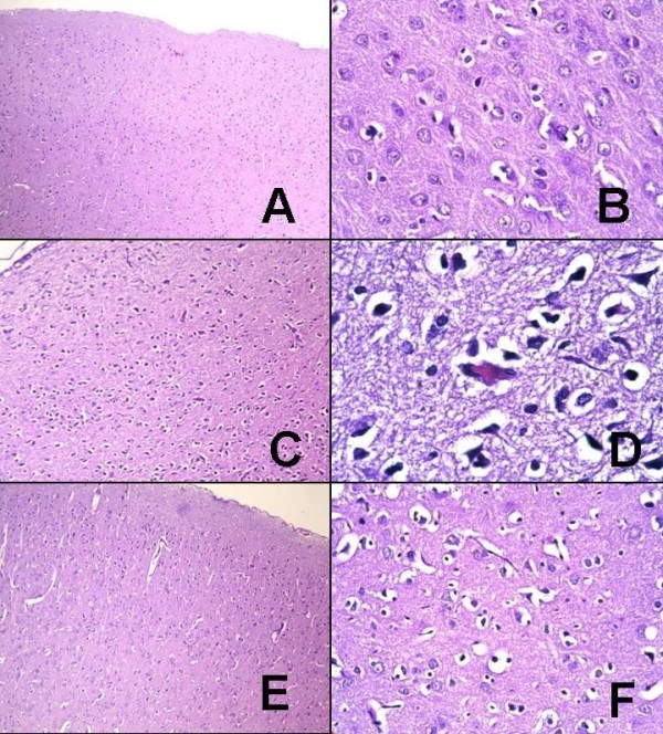
Figure 1.

Histological appearances of the groups. A, B: G1; histological appearances of normal brain parenchyma (hematoxylin and eosin (H&E), A: × 100, B: × 400). C, D: G2; edema in brain parenchyma, pyknotic changes (green arrow), nuclear hyperchromasia, and cytoplasmic eosinophilia (red arrow) in neurons are observed (H&E, C: × 100; D: × 1000, oil immersion). E, F: G4; axonal edema and moderate reactive and degenerative changes in neuronal structures are seen (H&E, E: × 100; F: × 400).
